Tumor Paint® Technology & CANVAS® Imaging
Blaze Bioscience has developed an investigational imaging agent and a dedicated near-infrared imaging system, working together to give surgeons real-time visibility of tumor tissue in the operating room.
Tozuleristide is an investigational imaging agent built on Blaze's Tumor Paint® technology. Its active component is a cystine-dense mini-protein (CDP), a chlorotoxin variant, conjugated to a near-infrared fluorescent dye. When administered intravenously before surgery, it exploits a set of molecular characteristics specific to cancer cells to accumulate selectively in tumor tissue.
Step 1: Surface binding. The CDP binds to molecular targets, including calpactin and MMP-2, that are clustered in lipid rafts on the surface of cancer cells, but are absent or less accessible on normal tissue.
Step 2: Trojan Horse entry. The cell pulls the CDP inside through clathrin-mediated endocytosis, a normal cellular uptake process. The fluorescent dye cargo comes with it, concealed like a Trojan Horse within the internalization pathway.
Step 3: Accumulation. Once inside, the CDP is retained and accumulates within the cancer cell. The result: tumor tissue becomes fluorescent and distinctly visible under near-infrared imaging while healthy tissue remains dark.
This mechanism of selective binding, active internalization, and intracellular retention gives tozuleristide its sensitivity and specificity across tumor types, and underpins its activity in five surgical indications studied to date.
Near-infrared and visible light overlay of a lumpectomy specimen reveals tozuleristide fluorescence during breast cancer surgery.
Intraoperative imaging, Phase 1 breast cancer study. Images on file, Blaze Bioscience. Investigational use only.
The CANVAS® imaging system is the intraoperative platform through which surgeons visualize the fluorescence signal produced by tozuleristide. Developed by Blaze Bioscience, the system has two components: a near-infrared imaging head that attaches to the operating microscope, and a dedicated imaging station that processes and displays the signal on an operating room monitor.
During surgery, CANVAS® delivers near-infrared illumination to the surgical field through the existing microscope optics. Tissue where tozuleristide has accumulated emits a fluorescence signal clearly distinct from the surrounding healthy tissue, providing a real-time visual guide to tumor boundaries without interrupting the operative workflow.
The system has been used across multiple brain cancer clinical studies, building an operational track record alongside the clinical data for tozuleristide itself.
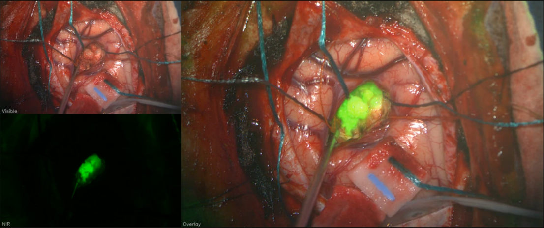
CANVAS® intraoperative imaging: visible light, near-infrared, and overlay views during brain tumor resection. Tumor tissue illuminated with tozuleristide.
Tozuleristide and CANVAS® are designed as a combined system. Together, they create an intraoperative workflow that gives surgeons a fluorescence-guided view of tumor tissue in real time.
Tozuleristide is administered intravenously before surgery. The agent distributes through the body and begins accumulating in cancer cells.
Tozuleristide internalizes preferentially within cancer cells, concentrating the near-infrared dye in tumor tissue while healthy tissue remains largely clear.
In the operating room, CANVAS® illuminates the surgical field with near-infrared light. Tumor tissue emits a visible fluorescence signal; healthy tissue does not.
Surgeons see a real-time fluorescence image of the tumor tissue, providing a visual guide to tumor boundaries at the critical moment of resection.
Blaze is now focused on breast cancer, advancing tozuleristide and CANVAS® to address one of the most critical unmet needs in breast-conserving surgery: real-time margin assessment.
Clinical Program Contact Us